Actinic Keratosis: Why Early Intervention Saves Skin Health
Actinic Keratosis Risk Assessment Tool
Which describes your skin best?
How much time do you spend outdoors?
What is your age range?
Do you have any of these conditions?
Quick Takeaways
- actinic keratosis is a sun‑induced precancerous skin lesion that can become squamous cell carcinoma.
- Early detection and treatment dramatically lower the risk of cancer and reduce scarring.
- Topical medications, cryotherapy, and photodynamic therapy all achieve cure rates above 80% when used promptly.
- Regular skin checks with a dermatologist are key for people with high sun exposure or a history of lesions.
- Simple sun‑protection habits-broad‑spectrum sunscreen, hats, and avoiding peak UV hours-prevent most new lesions.
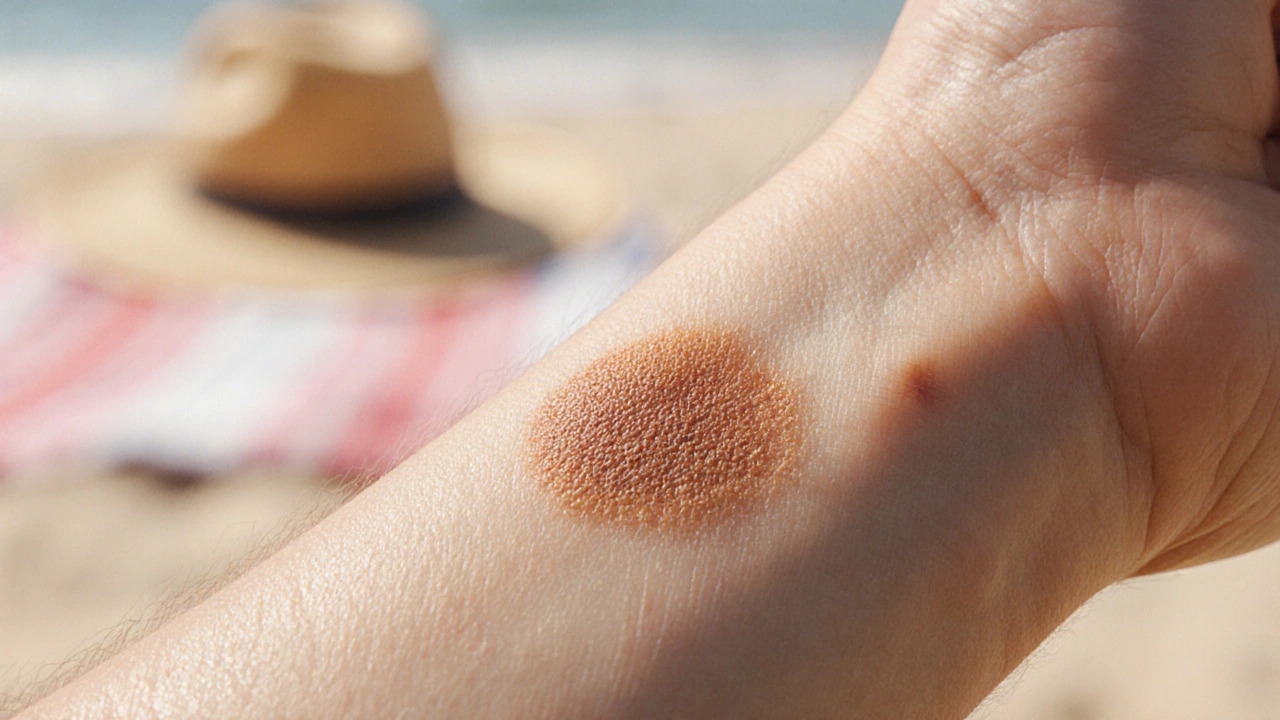
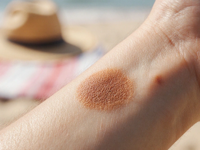
When you hear Actinic Keratosis is a rough, scaly patch that forms on sun‑exposed skin, usually after years of UV damage. Also called solar keratosis, it’s considered a precancerous lesion that can turn into squamous cell carcinoma if left unchecked.
What is Actinic Keratosis?
In plain terms, actinic keratosis (AK) looks like a tiny, sandpaper‑like bump or a reddish‑brown patch. They typically appear on the face, ears, scalp, forearms, or back of the hands-areas that soak up the most sun. While most AKs are painless, they can itch or bleed if scraped.
From a medical perspective, AK represents abnormal growth of keratinocytes caused by DNA damage from ultraviolet (UV) radiation. The damage accumulates over decades, and the body's repair mechanisms eventually fail, leading to these atypical cells.
Because AK sits on the spectrum between normal skin and invasive Squamous Cell Carcinoma (a type of skin cancer that can spread if untreated), it’s a red flag that the skin is vulnerable.
Why Early Intervention Matters
Statistics from the American Academy of Dermatology show that about one in three people over 60 has at least one AK. Without treatment, an individual lesion has a 0.025%‑0.5% yearly chance of becoming invasive SCC. The risk climbs sharply when multiple lesions are present or when the patient is immunosuppressed.
Acting early cuts that risk dramatically. Studies published in the Journal of Dermatologic Surgery (2023) found that lesions removed within six months of detection had a 99% clearance rate and virtually no progression to cancer.
Besides cancer avoidance, early treatment means simpler procedures. Small, isolated AKs respond well to cryotherapy or a short course of topical cream, whereas larger or long‑standing lesions may need surgical excision, which carries higher costs and potential scarring.
Who Is Most at Risk?
Age and Skin Type: Fair‑skinned individuals (Fitzpatrick types I‑II) accumulate UV damage faster. People over 50 have the highest prevalence.
Sun Exposure (cumulative ultraviolet radiation from outdoor activities) is the biggest driver. Outdoor workers, avid gardeners, and frequent beachgoers are especially vulnerable.
Other risk factors include:
- History of sunburns, especially in childhood.
- Chronic immunosuppression (organ‑transplant patients, long‑term steroids).
- Previous skin cancers-having one skin cancer raises the chance of future lesions.
- Genetic conditions like xeroderma pigmentosum.
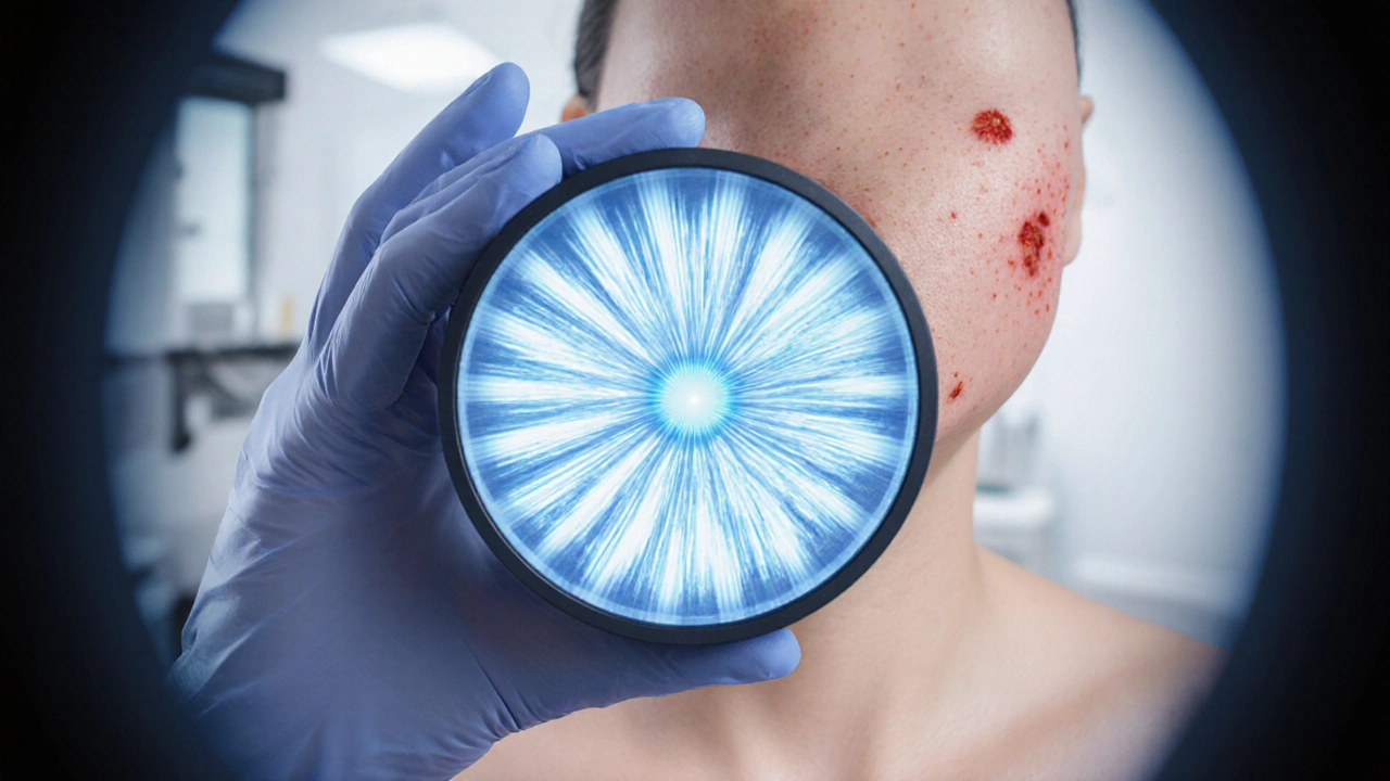
How Is Actinic Keratosis Diagnosed?
Diagnosis starts with a visual exam. Dermatologists use a dermatoscope-a magnifying device with polarized light-to spot characteristic patterns (the so‑called “strawberry” or “spoke‑wheel” appearance).
If the lesion looks atypical or if there are many lesions, a Biopsy (removal of a small skin sample for microscopic analysis) may be performed. Shave or punch biopsies confirm whether the cells are dysplastic or already cancerous.
Advanced imaging, such as reflectance confocal microscopy, can provide a non‑invasive view, but it’s rarely needed for routine AK work‑ups.
Treatment Options
Because AKs are numerous and often appear in clusters, clinicians tailor therapy to the lesion’s size, location, and the patient’s overall health. Below is a quick comparison of the most common approaches.
| Treatment | How it Works | Typical Sessions | Pros | Cons |
|---|---|---|---|---|
| Cryotherapy | Freezes lesion with liquid nitrogen, causing cell death. | 1‑2 per lesion | Fast, inexpensive, high cure rate for isolated AKs. | May cause blistering; less effective on thick lesions. |
| 5‑Fluorouracil (5‑FU) | Chemical that disrupts DNA synthesis in abnormal cells. | 2‑4 weeks of twice‑daily application | Treats large fields of AKs; visible cosmetic outcome. | Redness, crusting, and pain during treatment. |
| Imiquimod | Stimulates immune response to destroy dysplastic cells. | 2‑3 weeks per course, repeated up to 4 cycles | Good for immunocompromised patients; field therapy. | Local inflammation, flu‑like symptoms possible. |
| Photodynamic Therapy (PDT) | Photosensitizing cream applied then activated by blue/red light. | 1‑2 sessions, a week apart | Effective for many AKs at once; minimal scarring. | Requires clinic visit; temporary redness and swelling. |
| Surgical Excision | Physical removal of lesion with margin, followed by stitching. | Single procedure per lesion | Definitive removal, histologic confirmation. | Higher cost, possible scar, usually reserved for suspicious AKs. |
For most patients, a combination of field therapy (like 5‑FU or PDT) plus spot treatment (cryotherapy) yields the best result. Your dermatologist will decide based on lesion count, location, and skin type.
Follow‑up Care and Prevention
After treatment, the skin may look raw for a week or two. Keep the area clean, avoid harsh scrubs, and apply a fragrance‑free moisturizer.
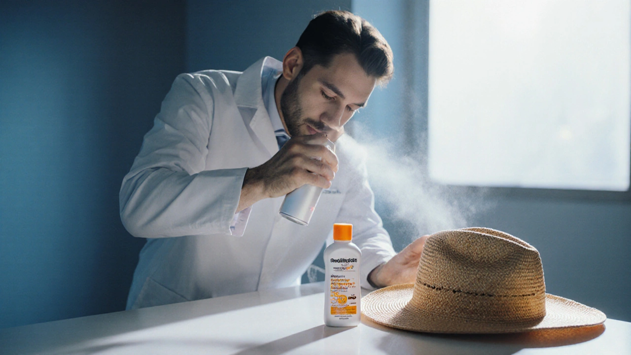
Long‑term prevention hinges on sun protection:
- Apply broad‑spectrum SPF30+ sunscreen every morning, reapply every two hours outdoors.
- Wear wide‑brim hats, UV‑blocking sunglasses, and long sleeves when the sun is strong.
- Seek shade between 10a.m. and 4p.m., when UV intensity peaks.
- Consider antioxidant‑rich skin care (vitaminC, niacinamide) to support DNA repair.
Schedule a full‑body skin exam with a Dermatologist (a skin‑care specialist trained to diagnose and treat skin conditions) at least once a year, or more often if you have a history of AKs.
Common Myths Debunked
Myth 1: “If it doesn’t hurt, it’s harmless.”
Reality: AKs are often painless, yet they can still progress to cancer.
Myth 2: “Only sun‑bathers get AK.”
Reality: Cumulative lifetime UV exposure matters more than occasional beach trips.
Myth 3: “A single treatment cures everything.”
Reality: Field‑therapy addresses hidden lesions that aren’t visible to the eye.
When to See a Dermatologist Right Away
Contact a dermatologist promptly if you notice any of the following:
- A lesion that bleeds, ulcerates, or grows rapidly.
- Persistent redness or a sore that won’t heal after two weeks.
- Multiple new AKs appearing within a short period.
- Any skin change on a scar, mole, or previously treated area.
Quick Action Checklist
- Perform a self‑skin exam monthly; note any new rough patches.
- Schedule a dermatologist visit if you find anything suspicious.
- Ask about field therapy if you have many lesions.
- Adopt daily sunscreen and protective clothing habits.
- Maintain follow‑up appointments every 6‑12 months.
Remember, catching actinic keratosis early isn’t just about avoiding a scar-it’s a proactive step toward preventing skin cancer. By staying vigilant and treating lesions promptly, you keep your skin healthier for years to come.
Frequently Asked Questions
Can actinic keratosis disappear on its own?
Rarely. While a few small lesions may regress, most persist or progress. Professional treatment is recommended for any confirmed AK.
Is cryotherapy painful?
You feel a brief, intense cold that numbs the area. After the freeze, a mild stinging sensation may occur for a day or two, but most patients tolerate it well.
How long does it take for topical creams to work?
Treatments like 5‑fluorouracil or imiquimod are applied for 2‑4 weeks. Visible improvement usually appears during the last week, with full clearance by the end of the regimen.
Should I stop using sunscreen after treatment?
No. Sun protection is crucial both during and after therapy to prevent new lesions and to aid healing of treated skin.
Is actinic keratosis a sign of other skin cancers?
AK indicates significant UV damage, which also raises the risk for basal cell carcinoma and squamous cell carcinoma. Regular skin checks are essential.
Jason Montgomery
Hey folks, just wanted to say that catching actinic keratosis early is like getting a tiny warning light before the car breaks down – it’s way easier (and cheaper) to fix it now than waiting for full‑blown skin cancer later. Grab that sunscreen, get regular skin checks, and don’t ignore those scaly patches. 🙌
Wade Developer
The early detection of actinic keratosis represents a quintessential exercise in preventive medicine.
The philosophical standpoint embodies the principle that small, observable deviations can preempt larger systemic failures.
In the dermatological realm, these lesions are not mere cosmetic nuisances but precursors with quantifiable malignant potential.
Epidemiological studies consistently demonstrate that timely excision or topical therapy reduces progression rates dramatically.
Clinicians therefore bear a duty to educate patients about the subtle visual cues that betray ultraviolet damage.
Patients, in turn, must cultivate a habit of self‑surveillance, examining their skin with the same diligence they would inspect a vital instrument.
The risk assessment tool operationalizes variables such as skin type, cumulative sun exposure, and immunological status into a coherent metric.
By translating these factors into a percentage, the tool demystifies abstract risk and renders it actionable.
Moreover, the stratified recommendations-from sunscreen reinforcement to biopsy-provide a clear therapeutic pathway.
It is worth noting that the underlying biology involves DNA photoproducts, oxidative stress, and impaired repair mechanisms.
These molecular cascades, once set in motion, can be halted or redirected only if interventions occur before clonal expansion.
Consequently, the financial and emotional burdens associated with advanced skin cancer are markedly attenuated by early action.
Public health policies that promote regular skin examinations parallel the broader ethos of anticipatory care.
In sum, the adage ‘prevention is better than cure’ finds a concrete illustration in the management of actinic keratosis.
Thus, embracing early intervention is not merely a recommendation but an ethical imperative for both providers and patients.
Sandra Perkins
Oh great, another reminder that “sunburns are fun” until you get a little wart.
rama andika
Yeah, because the sunscreen industry definitely isn’t making a fortune off our fear, right?
They’ve hidden a secret formula that only works if you believe in the conspiracy.
Meanwhile, the “big pharma” of dermatology pushes creams that cost more than a latte a day.
It’s all a grand ploy to keep us buying gadgets instead of actually staying out of the sun.
But hey, if you spot a flaky patch, just blame the government and move on.
Kenny ANTOINE-EDOUARD
For anyone wondering about treatment options, there are several evidence‑based pathways. Cryotherapy is quick and effective for isolated lesions. Topical agents like 5‑fluorouracil or imiquimod work well for field‑directed therapy when multiple spots are present. Photodynamic therapy offers a middle ground with good cosmetic outcomes. It’s important to discuss with a dermatologist which method aligns with your lesion count, skin type, and tolerance for side effects. Early intervention also allows for simpler procedures, reducing scar risk and recovery time.
Craig Jordan
Honestly, I find it amusing how often people quote guidelines without ever considering individual variability.
Sure, cryotherapy sounds “simple,” but have you thought about the pain tolerance of the average patient?
And those topical creams? They demand weeks of strict adherence, which most folks simply can’t maintain.
Photodynamic therapy may look glamorous, yet the cost and need for specialized equipment make it inaccessible for many.
What’s often omitted is the psychosocial impact-people may avoid treatment because of the stigma of visible skin changes.
Moreover, the literature you cite tends to cherry‑pick studies with industry funding.
In practice, a balanced approach that weighs patient preference, lesion biology, and realistic follow‑up capacity is far more prudent.
So before you push a one‑size‑fits‑all regimen, remember that dermatology is as much an art as a science.
Jeff Quihuis-Bell
Listen up, squad! 🚀 The best offense against actinic keratosis is a daily sunscreen habit and regular skin checks – treat it like a morning workout for your face.
If you spot anything weird, don’t play hero; book that derm appointment ASAP.
Remember, the sooner you act, the less dramatic the treatment will be – and you’ll keep that glow without the drama of surgery.
Jessica Tang
I agree with the emphasis on early detection, and I’d add that educating family members, especially older relatives, can amplify the preventive effect.
Sharing resources like the risk assessment tool helps demystify the process and encourages proactive skin exams at home.
Tracy Winn
Wow, what a thorough rundown – really covers everything from sunscreen to surgery, and it’s all so easy to follow! 🎉
Jessica Wheeler
While the enthusiasm is appreciated, it’s crucial to remember that neglecting the underlying lifestyle factors-like chronic tanning-undermines any topical solution. A disciplined approach to sun avoidance should be the foundation before any cream or procedure is considered.
Mikayla Blum
Exactly, staying out of the sun is key 😎